paper-plane envelope home office pencil quill pen image images camera play bullhorn connection mic file-text2 file-picture file-music file-play file-video copy folder folder-open folder-plus folder-minus folder-download folder-upload price-tag price-tags ticket phone envelop pushpin location compass map map2 clock alarm fax mobile bubble bubbles user users user-plus user-minus user-check quotes-left quotes-right search pie-chart stats-dots stats-bars airplane cloud-download cloud-upload earth link flag eye eye-blocked arrow-up-left arrow-up arrow-up-right arrow-right arrow-down-right arrow-down arrow-down-left arrow-left2 share amazon google-plus google-drive facebook instagram twitter rss youtube flickr dropbox linkedin file-pdf file-openoffice file-word file-excel
XClose

HiP-CT
Hierarchical Phase-Contrast Tomography (HiP-CT)

Home
Menu

Publications

Publication highlights:

Imaging intact human organs with local resolution of cellular structures using hierarchical phase-contrast tomography

Authors: C. L. Walsh, et al.
Journal: Nature Methods
DOI: 10.1038/s41592-021-01317-x
This paper presents a new synchrotron x-ray imaging technique, called Hierarchical Phase-Contrast Tomography (HiP-CT), which is used to span a previously poorly explored scale in our understanding of human anatomy, the micron to whole intact organ scale.
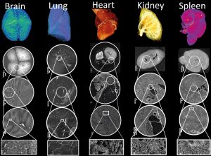
Human Organ AtlasHuman Organ Atlas, see videos at Gallery

The bronchial circulation in Covid-19 pneumonia

Authors: M. Ackermann, et al.
Journal: American Thoracic Society
DOI10.1164/rccm.202103-0594IM
This paper shows how COVID-19 disrupts the blood vessel network architecture of the lung, specifically we show cases of bronchio-pulmonary shunting in intact COVID-19 lung lobes.
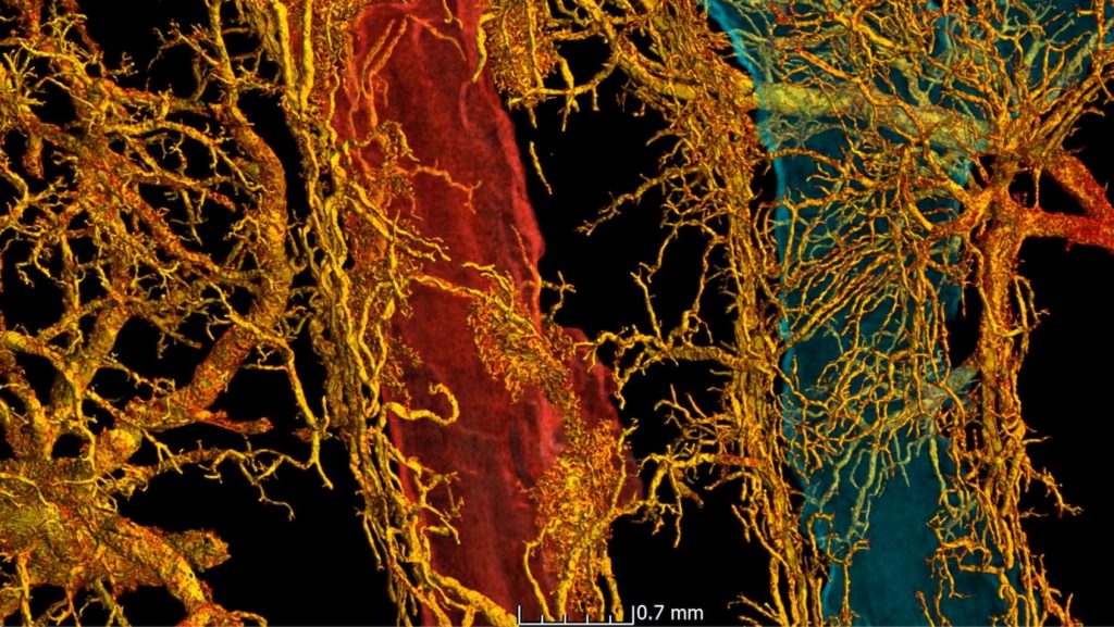
Bronchio-pulmonary shunting in a SARS-CoV-2 infected lungBronchio-pulmonary shunting in a SARS-CoV-2 infected lung

The fatal trajectory of pulmonary COVID-19 is driven by lobular ischemia and fibrotic remodelling

Authors: M. Ackermann, et al.
Journal: eBioMedicine
DOI10.1016/j.ebiom.2022.104296
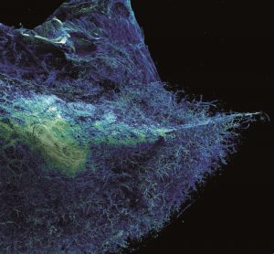
This paper identifies a link between the damage that severe Covid-19 can inflict on lungs and pulmonary fibrosis, a disease that causes severe scarring of lung tissue.
All Publications

    1. Walsh, C. L., et al. “Imaging intact human organs with local resolution of cellular structures using hierarchical phase-contrast tomography.” Nature methods 18.12 (2021): 1532-1541. DOI: 10.1038/s41592-021-01317-x
    2. Ackermann, M., et al. “The bronchial circulation in COVID-19 pneumonia.” American Journal of Respiratory and Critical Care Medicine 205.1 (2022): 121-125. DOI: 10.1164/rccm.202103-0594IM
    3. Xian, R.P., et al. “A multiscale X-ray phase-contrast tomography dataset of a whole human left lung.” Scientific Data 9, 264 (2022). DOI: 10.1038/s41597-022-01353-y
    4. Jonigk, D., et al. “Pulmonary and Systemic Pathology in COVID-19: Holistic Pathological Analyses.” Dtsch Arztebl Int 119 (2022): 429–35. DOI: 10.3238/arz-tebl.m2022.0231 (German) 10.3238/arztebl.m2022.0231 (English)
    5. Ackermann, M., et al. “The fatal trajectory of pulmonary COVID-19 is driven by lobular ischemia and fibrotic remodelling.” EBioMedicine 85 (2022): 104296. DOI: 10.1016/j.ebiom.2022.104296
    6. Brunet, J., et al. “Preparation of large biological samples for high-resolution, hierarchical, synchrotron phase-contrast tomography with multimodal imaging compatibility.” Nature protocols (2023): 1-27.  DOI: 10.1038/s41596-023-00804-z
    7. Bellier, Alexandre, et al. “Imagerie en contraste de phase par source synchrotron appliquée à l’étude anatomique de la vascularisation artérielle de l’hippocampe.” Morphologie 106.354 (2022): S22-S23. DOI: 10.1016/j.morpho.2022.06.018 (French)
    8. Bellier, Alexandre, et al. “Anatomie multi-échelle par imagerie synchrotron de la vascularisation bronchique dans l’infection à SARS-CoV-2.” Morphologie 106.354 (2022): S38. DOI: 10.1016/j.morpho.2022.06.051 (French)
    9. Ackermann, Maximilian, et al. “Reply to: Intrapulmonary Shunt and Alveolar Dead Space in a Cohort of Patients with Acute COVID-19 Pneumonitis and Early Recovery.” The European respiratory journal: 2202121. (2022). DOI: 10.1183/13993003.02121-2022
    10. Ackermann, Maximilian. “Auf dem Weg zum virtuellen Mikroskop: Synchrotron-basierte dreidimensionale Bildgebung in der Pathologie.” Die Pathologie (2022): 1-6. DOI: 10.1007/s00292-022-01161-6 (German)
    11. Kamp, Jan C., et al. “Novel Insight into Pulmonary Fibrosis and Long COVID.” American Journal of Respiratory and Critical Care Medicine 207.8 (2023): 1105-1107. DOI: 10.1164/rccm.202212-2314LE 
    12. Jain, Yashvardhan, et al. “SenNet + HOA – Hacking the Human Vasculature in 3D.” Kaggle. https://kaggle.com/competitions/blood-vessel-segmentation
    13. Xian, R. Patrick, et al. “A closer look at high-energy X-ray-induced bubble formation during soft tissue imaging.” Journal of Synchrotron Radiation 31.3 (2024): 566-577. DOI: 10.1107/S160057752400290X
    14. Lee, Ju Young, et al. “Protocol for 3D virtual histology of unstained human brain tissue using synchrotron radiation phase-contrast microtomography.” Frontiers in Physics 11 (2024): 1335285. DOI: 10.3389/fphy.2023.1335285
    15. Brunet, Joseph, et al. “Multidimensional Analysis of the Adult Human Heart in Health and Disease Using Hierarchical Phase-Contrast Tomography.” Radiology 312.1 (2024): e232731. DOI: 10.1148/radiol.232731
    16. Verleden, Stijn E., et al. “Imaging Three-Dimensional Airway Morphology in Congenital Pulmonary Airway Malformation Using Hierarchical Phase Contrast Tomography.” American Journal of Respiratory and Critical Care Medicine ja (2024). DOI: 10.1164/rccm.202403-0509IM
    17. Bellier, Alexandre, et al. “Micro to macro scale anatomical analysis of the human hippocampal arteries with synchrotron hierarchical phase-contrast tomography.” Surgical and Radiologic Anatomy (2024): 1-8. DOI: 10.1007/s00276-024-03467-x
    18. Submitted: Rahmani, Shahrokh, et al. “Micro to macro scale analysis of the intact human renal arterial tree with Synchrotron Tomography.” (March 2023) DOI: 10.1101/2023.03.28.534566
    19. Submitted: Brunet, Joseph, et al. “Hierarchical Phase-Contrast Tomography: Advancing High-Resolution Imaging of Intact Human Organs.” (October 2023), Gray’s Anatomy.
    20. Submitted: Yagis, Ekin, et al. “Deep Learning for Vascular Segmentation and Applications in Phase Contrast Tomography Imaging.” (November 2023) DOI: 10.48550/arXiv.2311.13319
    21. Submitted: Mirone, Alessandro,  et al. “Eikonal phase retrieval: Unleashing the fourth generation sources potential for enhanced propagation based tomography on biological samples.” (November 2023) DOI: 10.48550/arXiv.2311.14745
    22. Submitted: Parmenter, Alissa, et al. “Multimodal imaging reveals multiscale mechanical interplay in vertebral endplate microarchitecture during intervertebral disc loading” (August 2024) DOI: 10.1101/2024.08.19.608559
    23. Submitted: Jain, Yashvardhan, et al. “Vasculature segmentation in 3D hierarchical phase-contrast tomography images of human kidneys” (August 2024) DOI: 10.1101/2024.08.25.609595
Back to top