Publication highlights:
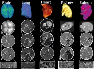
Imaging intact human organs with local resolution of cellular structures using hierarchical phase-contrast tomography
Authors: C. L. Walsh, et al.
Journal: Nature Methods
DOI: 10.1038/s41592-021-01317-x
This paper presents a new synchrotron x-ray imaging technique, called Hierarchical Phase-Contrast Tomography (HiP-CT), which is used to span a previously poorly explored scale in our understanding of human anatomy, the micron to whole intact organ scale.
 Human Organ Atlas, see videos at Gallery
The bronchial circulation in Covid-19 pneumonia
Authors: M. Ackermann, et al.
Journal: American Thoracic Society
DOI: 10.1164/rccm.202103-0594IM
This paper shows how COVID-19 disrupts the blood vessel network architecture of the lung, specifically we show cases of bronchio-pulmonary shunting in intact COVID-19 lung lobes.
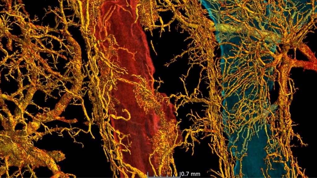 Bronchio-pulmonary shunting in a SARS-CoV-2 infected lung
The fatal trajectory of pulmonary COVID-19 is driven by lobular ischemia and fibrotic remodelling
Authors: M. Ackermann, et al.
Journal: eBioMedicine
DOI: 10.1016/j.ebiom.2022.104296
This paper identifies a link between the damage that severe Covid-19 can inflict on lungs and pulmonary fibrosis, a disease that causes severe scarring of lung tissue.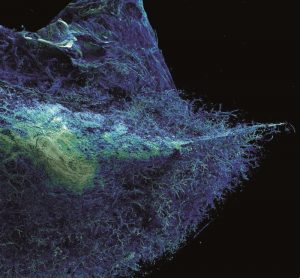
 Close
Close
