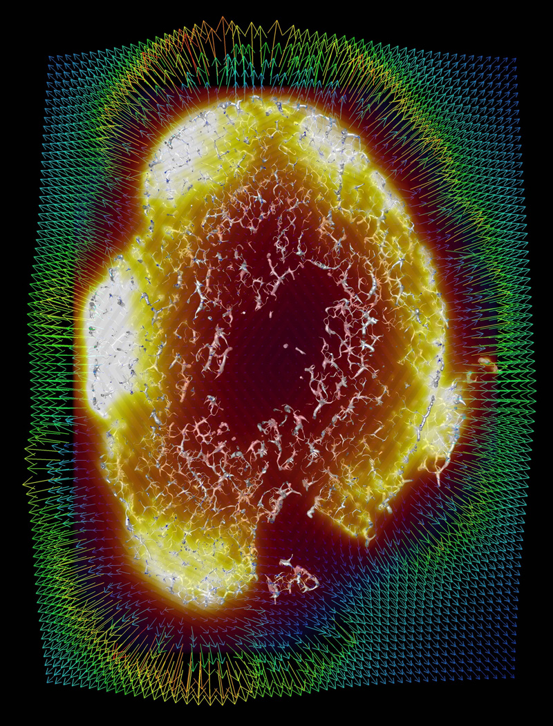
Imaging of cleared tissue and modelled flow to predict drug uptake and treatment responses in tumours. See d’Esposito et al, Nature Biomedical Engineering, 2, 773-787 (2018). (Credit S. Walker-Samuel)
The Hip-CT project arose from the desire to image the cellular level injury in Covid-19 victim lungs.
Early in the pandemic a group of medics at Mainz and Hannover contacted Professor Peter Lee at UCL to see if synchrotron X-ray imaging could be used to quantify the injury Covid-19 causes in the lung. We quickly brought together scientists at ESRF and Diamond Light Source and started imaging biopsy (tiny) samples.
Paul Tafforeau at ESRF proposed that using the ESRF-EBS we could get the same resolution on whole organs as we do in biopsies, which would be transformational as we could link cellular to organ scale damage. Forming a UK-French-German team, we developed HiP-CT during lockdown, and we are continuing to develop it, while using the results to help clinicians understand the injury Covid-19 has on our organs.
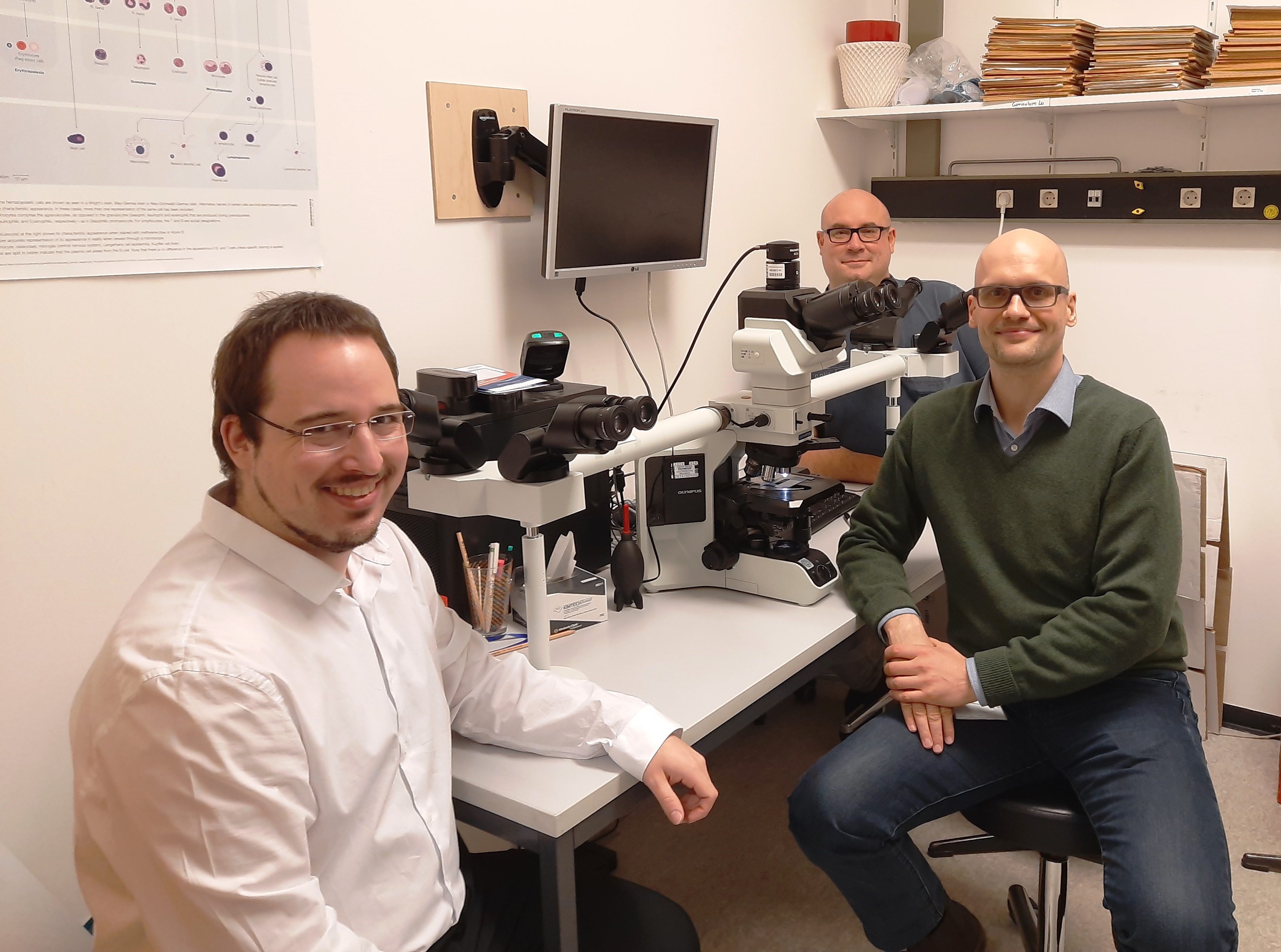
The team of Hannover Medics work on correlating the HiP-CT imaging down to sub-cellular and genetic levels. Danny Jonigk, Christopher Werlein and Mark Kuehnel
 Close
Close
