What is Hierarchical Phase-Contrast Tomography (HiP-CT)?

Prof Peter Lee, Drs Claire Walsh and Paul Tafforeau imaging the complete brain of a Covid-19 victim using HiP-CT , resolving cellular features (ca. one-micron resolution) in local areas.
Hierarchical Phase-Contrast Tomography (HiP-CT) is an X-ray technique which promises to decouple field of view and resolution to image anywhere in the human body with cellular (micron) resolution. The technique will enable the scanning of whole bodies with a resolution of 25 microns, thinner than a human hair and ten times the resolution of a medical CT scanner. Areas can then be selected to be zoomed in on, achieving local micron resolution, or one hundred times the resolution of medical CT.
Funded by the Chan Zuckerberg Initiative (CZI), the project is an international interdisciplinary collaboration between scientists and mathematicians at University College London (UCL), European Synchrotron Radiation Facility (ESRF) and Diamond Light Source (DLS), and clinicians at Hannover-biobank, Mainz, Heidelberg, UCL and Imperial College London, together with many other collaborators. The imaging advances are based on the recent Extremely Brilliant Source (EBS) upgrade to the ESRF that has created the world’s first high-energy fourth-generation synchrotron, which is currently the brightest X-ray source in the world. Feasibility studies have already demonstrated it can resolve unprecedented detail revealing the damage caused by Covid-19 on human lungs, linking from the major airways all the way down to the finest micro-vasculature in an intact lung, as shown in the video below.
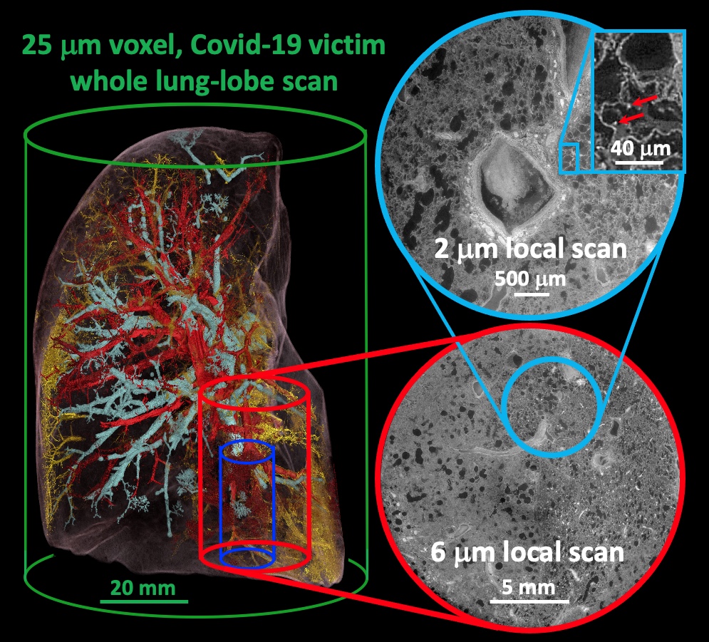
HiP-CT characterisation of the damage in a Covid-19 injured lung lobe zoom from the whole organ down to 2 micron resolution.
 Close
Close
